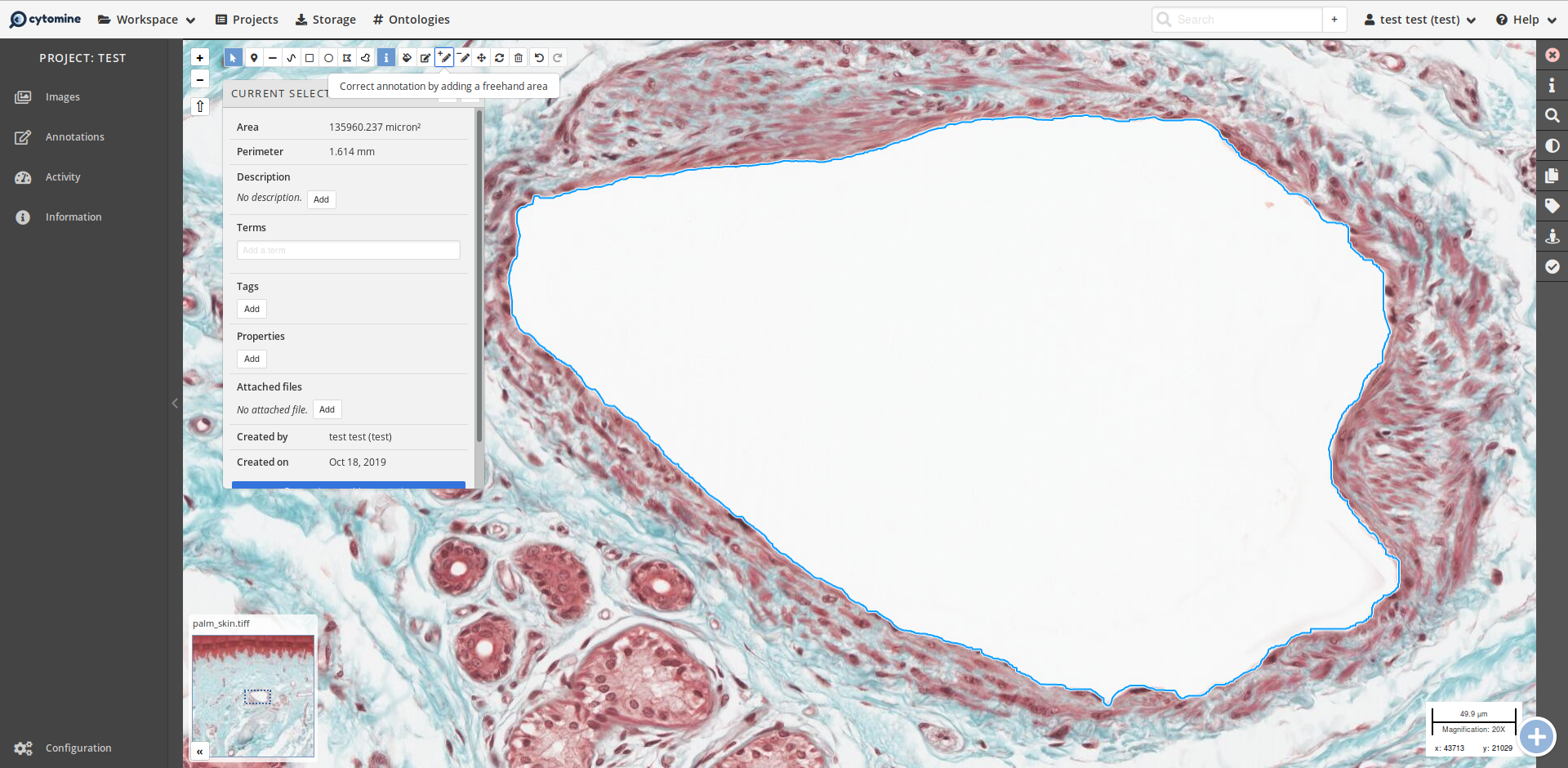
Image annotation and bio-image database

Guillaume Gay, CENTURI

June 10 2021

We are here to talk about microscopy image databases. We are not going to talk a lot about the “database” part of that, because a lot has to be said about microscopy and images before that is more important.

Follow this course here:

centuri-engineering.univ-amu.fr/BioImageDB/

Why you should care?

An excel file does not consitute a database

Excel is an accounting tool
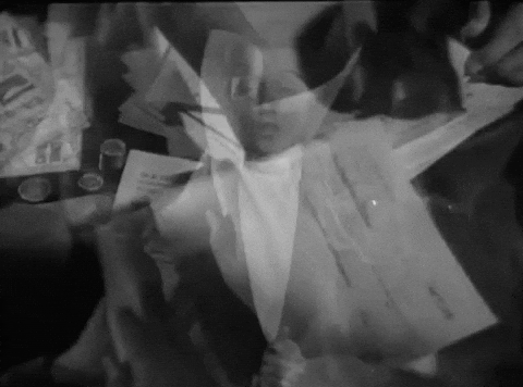
Course outline

  1. Some history of microscopy techniques

  2. The digital image (data and metadata)

  3. Databases (at last)

Some technique history

Early Microscopes

Antonie van Leewenhoek (1632–1723)

Robert Hooke (1635-1703)

HookeFlea01.jpg

National Library of Wales, Public Domain

First detector is the eye, data is registered through drawings.

Santiago Ramón y Cajal (1852 - 1934)

PurkinjeCell.jpg
Public Domain, Link

The eye & hand are still the best detector in the early XXth century.

First photos

Henry Fox Talbot (1800 - 1877)

Photomicrograph of insect wings - By William Henry Fox Talbot

See this article

First movies

Jean Comandon in 1909

Haemanthus katherinae (1956!)

Mitosis in Haemanthus katharinae endosperm

Technique evolution

Fluorescence !

  • dark field
  • multiple colors
  • specificity - we observe not only the organism but a precise molecule within the organism.

The confocal microscope

Davidovits & Egger 1969

  • The detector is a photomuliplier - first time the image from the microscope is a signal
  • Only the light emitted at the focal point is recorded.

Here comes the CCD

Some details on how it works here

  • Photon counting!

  • The image is a quantitative, digital, signal

From now on, an image is represented by a matrix of pixels

Modern microscopes

The super resolution revolution

Do you know Abbe law?

\[d = \frac {\lambda}{2 n A} \]

The minimum size of a motif - for exemple the distance between two spots, observable under a microscope is limited by the objective numerical aperture and the emission wavelength.

We invented ways to beat that limit!

(can you cite super resolution methods?)

An exemple: STORM

Christophe Leterrier, NeuroCyto, INP, Marseille

An other: Lattice light sheet

Movie 11 High Resolution from HHMI NEWS on Vimeo.

Sreens and plates

Multiple wells under a microscope on a moving stage

Conclusion

Image aquisition methods have always been immediatly applied to microscopy

The eye was surpassed only recently

The image became digital only 20 years ago!

The digital image

What is important to know?

Data and metadata

Can you cite image formats?

TIFF is the norm

TIFF is for Tagged Interchange File Format

A TIFF is a structured file with a header before the data:

The tiff file structure

We have tags to store metadata !

Tiff tags

What an 8 by 8 pixel file looks like:

An 8x8 pixels block
00000000: 4949 2a00 0800 0000 0e00 0001 0400 0100  II*.............
00000010: 0000 0800 0000 0101 0400 0100 0000 0800  ................
00000020: 0000 0201 0300 0100 0000 0800 0000 0301  ................
00000030: 0300 0100 0000 0100 0000 0601 0300 0100  ................
00000040: 0000 0100 0000 0e01 0200 1200 0000 b600  ................
00000050: 0000 1101 0400 0100 0000 3001 0000 1501  ..........0.....
00000060: 0300 0100 0000 0100 0000 1601 0400 0100  ................
00000070: 0000 0800 0000 1701 0400 0100 0000 4000  ..............@.
00000080: 0000 1a01 0500 0100 0000 0801 0000 1b01  ................
00000090: 0500 0100 0000 1001 0000 2801 0300 0100  ..........(.....
000000a0: 0000 0100 0000 3101 0200 0c00 0000 1801  ......1.........
000000b0: 0000 0000 0000 7b22 7368 6170 6522 3a20  ......{"shape":
000000c0: 5b38 2c20 385d 7d00 0000 0000 0000 0000  [8, 8]}.........
000000d0: 0000 0000 0000 0000 0000 0000 0000 0000  ................
000000e0: 0000 0000 0000 0000 0000 0000 0000 0000  ................
000000f0: 0000 0000 0000 0000 0000 0000 0000 0000  ................
00000100: 0000 0000 0000 0000 0100 0000 0100 0000  ................
00000110: 0100 0000 0100 0000 7469 6666 6669 6c65  ........tifffile
00000120: 2e70 7900 0000 0000 0000 0000 0000 0000  .py.............
00000130: 0101 0101 0101 0101 0101 0101 0101 0101  ................
00000140: 0101 0101 0101 0101 0101 0101 0101 0101  ................
00000150: 0101 0101 0101 0101 0101 0101 0101 0101  ................
00000160: 0101 0101 0101 0101 0101 0101 0101 0101  ................

A sad story

  • In the 90’s - 2000’s, MetaMorph software dominates the industry, has its own ‘format’
  • Eventually, constructors build their own software, try to impose it, how?

\(\Rightarrow\) Lots of incompatible & proprietary formats

OME to the rescue

«It is possible to interpret images only if we know the context in which they were acquired»

(Swedlow et al. 2003)

The OME-TIFF Format

We can put “things” in the TIFF header - so why not all the metadata we can think off?

This became a standard

The whole schema

Is available here

Look for the important points you thought of.

What do you think of XML?

In a file

<Image ID="Image:0" Name="Excy2_4.6.+12.lif [Excy2 4.6 - Phall CD24 Org 2]">
  <AcquisitionDate>2016-05-20T13:08:29
  </AcquisitionDate>
<ImagingEnvironment/>
<Pixels BigEndian="true"
        DimensionOrder="XYCZT"
        ID="Pixels:0"
        Interleaved="false"
        PhysicalSizeX="0.4814710371819961"
        PhysicalSizeXUnit="µm"
        PhysicalSizeY="0.4814710371819961"
        PhysicalSizeYUnit="µm"
        SignificantBits="8"
        SizeC="4"
        SizeT="1"
        SizeX="512"
        SizeY="512"
...

See more details here

Limits to OME-XML

What did you say a microscope image was?

The future: how to define flexible, “just general enough” file formats.

Let’s look at ZARR

The other metadata

But there’s more! The organism, the protocol, gene deletion,

too much!

Resort to ontologies

Global consortium QUAREP - LiMi

Finally Databases!

One DB system to rule them all: OMERO

We happen to have one here

The Contender Cytomine (but still using BioFormats!)

Cytomine is oriented towards collaboration after the image is produced.

Annotation in Cytomine

Public microscopy image databases

A word on FAIR

  • We need to be able to reuse data
  • We must be able to do this automatically

Published here

Findability

Accessibility

Interoperability

Reusability

A non exhaustive list:

The Allen Institute

Brain Atlases

See here

The Allen Cell explorer

  • Tries to know all the possible states of stem cells
The strategy
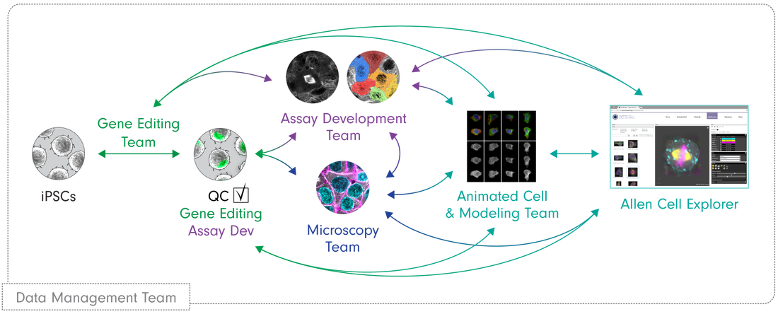
  • Created an extensive catalog of cell structures

4D Nucleome

A platform to search, visualize, and download nucleomics data.

Icludes microscopy data

European initiatives

Under the BioImage Archive

The Future ™

  • Lots of efforts towards FAIR - Fr, EU, Worldwide infrastructure
  • Crossing microscopy with other *omics data
  • Setting up standards is very hard

Conclusion

Already a lot of ressources but

  • Little actual re-use for now
  • Not used everywhere (biologists are still reluctant to share)
  • Please annotate your data
  • Talk to me if you need omero!
// reveal.js plugins